The first dental X-ray was taken way back in 1985. It was an X-ray of a molar tooth. Back then, the procedure took as long as 25 minutes! Today, digital X-rays have replaced older radiographic methods, emitting up to 80% less radiation than before. It is almost impossible to imagine modern dental diagnostics without X-rays. X-rays reveal those secrets about your oral health that a dentist’s naked eye cannot detect on its own!
Why is a dental X-ray necessary?
While it is possible to notice many changes in the oral cavity during an examination, some issues remain hidden and cannot be detected without additional diagnostics. A dental X-ray helps us identify various conditions, such as hidden cavities between teeth, diseases affecting the structures around the teeth and jawbones, the presence and positioning of tooth buds, abscesses, cysts, and even tumors.
Detecting these conditions early enables us to take timely action to prevent disease progression, pain, as well as extended treatment, which could lead to higher costs. This is why dental X-rays are essential for diagnostics, and the ultra-low doses of radiation you are exposed to during the procedure are negligible compared to the benefits of early detection and treatment.
What imaging method does Nova Dental Clinic use?
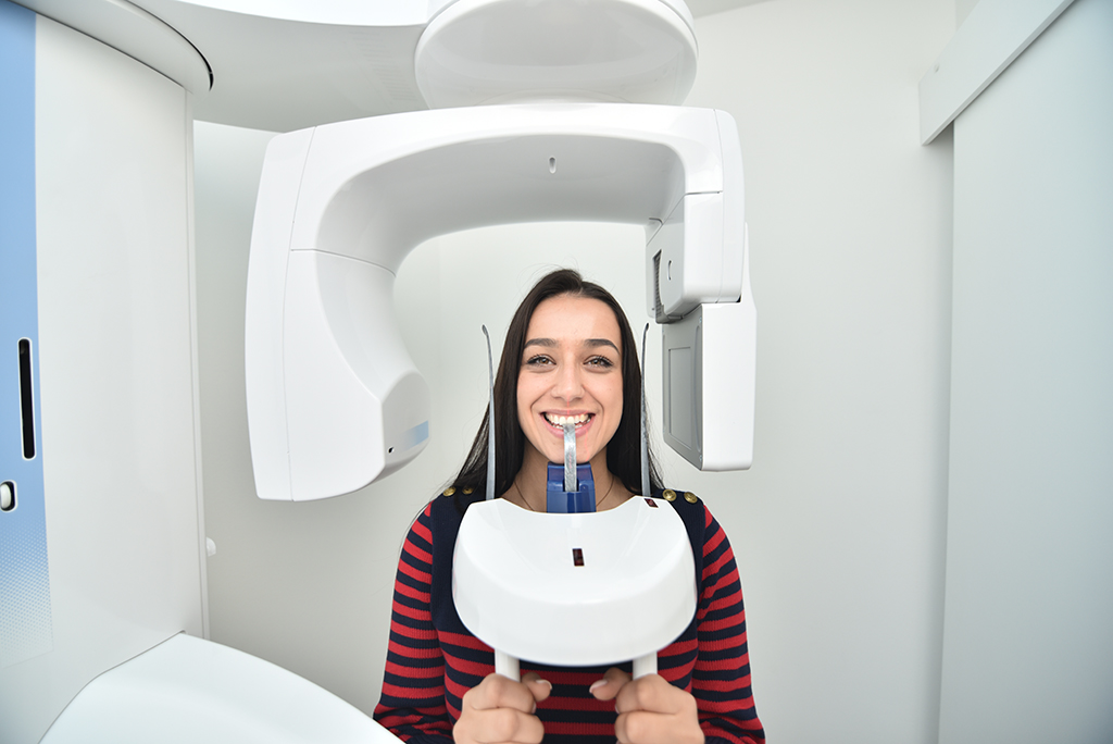
At Nova Dental Clinic, we use the Planmeca ProMax 3D Max digital imaging system for your X-rays. One of the most awarded devices in dentistry, it is renowned for its functionality, innovation, eco-friendliness, and design. Aside from being able to capture 2D images, this device also uses 3D imaging technology, allowing precise diagnostics and planning of dental procedures. This device employs Cone Beam Computed Tomography (CBCT) and digital radiography. Thanks to its sensors and advanced software algorithms, the Planmeca ProMax 3D Max ensures maximum accuracy with minimal risk for the patient.
The advantages of digital imaging over traditional X-rays are numerous, including lower radiation doses and a simplified imaging process. The ultra-low-dose minimizes the risk of harmful consequences of radiation exposure, while also making sure that the images are of high-quality. With ultra-low-dose technology, the imaging procedure is simplified, which is evident in its short duration.
The imaging with the Planmeca device lasts only about fifteen seconds.
It’s worth noting that diagnostics and treatment planning using these images can be applied across all fields of dentistry, including implantology, periodontology, endodontics, orthodontics, and oral surgery.

How to know which type of dental X-Ray you need??
Nova Dental Clinic offers several types of X-rays, depending on the condition of your teeth and surrounding structures, as well as the planned therapy.
These include:
- Panoramic X-Ray (Orthopantomogram or “OPG”) – A 2D image that provides an overview of the condition of your teeth and jaws, assisting in planning numerous treatments such as prosthetics, orthodontics, or periodontics.
- Panoramic Sinus X-Ray – Provides a 2D visualization of the sinuses aiming to detect potential changes or abnormalities.
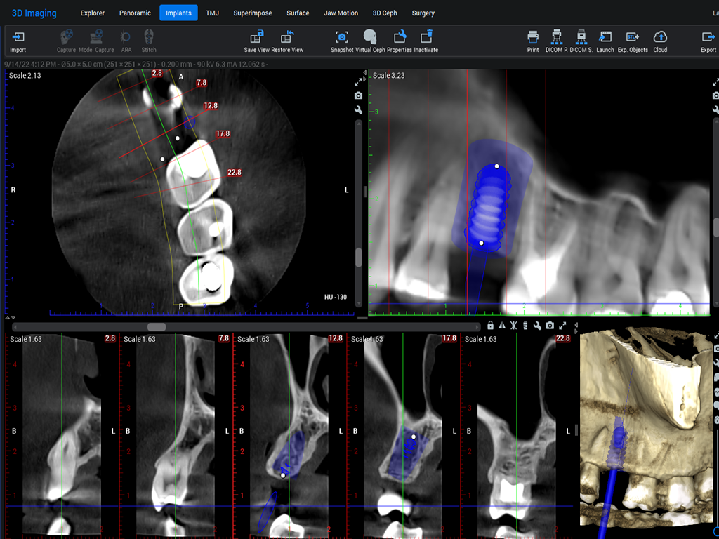
CBCT scans are available in various sizes depending on the teeth and jaw area of interest for diagnostics and therapy. Among the most commonly requested are:
- CBCT Scan of the Dental Region – 5×5 cm – For diagnosing the condition of individual teeth, their roots, root canals, and surrounding bone. Useful for endodontic treatment planning, evaluating the condition of the tooth before crown placement, implant placement, and wisdom tooth removal.
- CBCT Scan of One Jaw – 8×5 cm – In cases where the focus of the dental therapy is limited to either the upper or the lower jaw. It is used in cases where it is certain that diagnostics and therapy are required for a singular jaw. This type of scan can also capture the patient’s sinuses, as directed by a maxillofacial surgeon, in cases of suspected inflammatory changes in the sinus mucosa or the presence of cysts, as well as to determine the connection between the roots of posterior teeth and the sinus floor.
- CBCT Scan of Both Jaws – 8×8 cm – Covers all teeth and both jaws. Therapy planning and teeth rehabilitation involving both jaws indicates the necessity of this type of scan, by which we can evaluate the condition of the surrounding bone, which can be useful for periodontics and implantology.
- CBCT Scan of Both Jaws and the Surrounding Structures – “expanded field,” 11×8 cm – This “expanded field” imaging allows us to evaluate not only all the teeth but also areas beyond the last teeth in both jaws, aiding in implant placement and identifying unerupted teeth along with their connection to surrounding structures prior to surgical procedures.
When selecting the type of imaging as a diagnostic tool, we follow the so-called ALARA principle (As Low as Reasonably Achievable).
This means that we establish an indication for imaging for each patient, and determine individually which type of scan is necessary based on the condition in their oral cavity, and we also ensure that each patient is exposed to the lowest radiation dose necessary for their diagnosis.
Modern diagnostic methods have significantly advanced dental treatment planning, simplifying the treatment process for both doctors and patients. Poor oral health impacts overall well-being, that’s why we can consider oral care the first step toward a healthier life.
At Nova Dental Clinic, every new patient undergoes a detailed dental examination supported by orthopantomogram. For complex therapies and full jaw rehabilitations, 3D imaging analysis is indispensable before starting the treatment. To achieve a healthy and beautiful smile with top-notch diagnostics, schedule your appointment today!vna. Kako bismo i vama pružili zdrav i lijep osmijeh pomoću vrhunske dijagnostike, zakažite svoj termin već danas!